Pracownik naukowy w Instytucie Medycyny Regeneracyjnej McGowan na Uniwersytecie w Pittsburghu w stanie Pensylwania (USA) gdzie zajmuje się badaniami nad regeneracją tkanek.

Członek ISAKOS Leg, Ankle & Foot (LAF) Committee. (Międzynarodowe Towarzystwo Artroskopii, Chirurgii Kolana i Ortopedycznej Medycyny Sportowej)
Członek ESSKA Basic Science Committee. (Europejskie Towarzystwo Traumatologi Sportowej, Chirurgii Kolana i Artroskopii)
Recenzent w prestiżowym dwumiesięczniku naukowym wydawanym przez American College of Foot and Ankle Surgeons
Recenzent w renomowanym czasopiśmie naukowym Europejskiego Towarzystwa Traumatologii Sportowej, Chirurgii Kolana i Artroskopii
Współautorstwo rozdziałów w międzynarodowych podręcznikach i książkach z zakresu ortopedii i traumatologii sportowej.

Techniques for Treating the Hip, Knee, and Ankle
Abstract: Understanding meniscus anatomy is a key to success for meniscus surgical treatment. The focus of this chapter is to discuss the medial and lateral meniscus anatomy, including detailed description of its ligaments and connections to the femur, tibia, and fibula. This knowledge is essential while suturing or transplanting meniscus.
Open and Arthroscopic Techniques
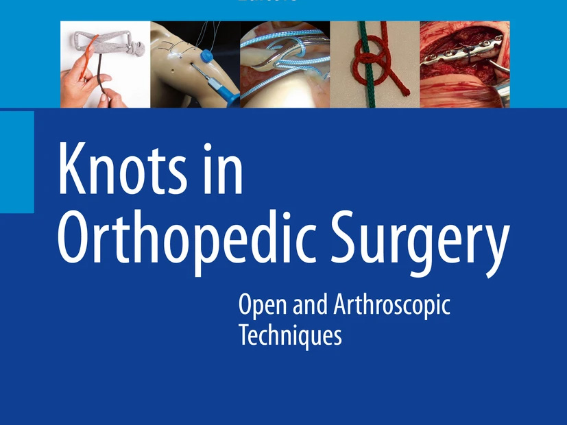
Abstract: Suture manipulation is a key point in arthroscopic surgery. Sutures are mainly used in shoulder surgeries. However, during meniscal repairs, some ankle and elbow arthroscopic procedure surgeons need ability of suture manipulation. In this chapter, we will focus on suture manipulations during shoulder arthroscopies such as basic suture handling and configurations. All these maneuvers can be adapted to other procedures in different joints.
An Evidence-Based Medicine Approach
Abstract: “Ribbon” concept of anterior cruciate ligament (ACL) anatomy has become one of the hottest topics in orthopedic world recently. In this chapter, we are going to present key points of this concept including direct and indirect insertion at the femoral side; flat, twisting nature of midsubstance of the ACL; and “C”-shaped appearance, with different anatomical variations on the tibial side. The chapter is accompanied by detailed anatomical dissection pictures.
A Comprehensive Review of their Anatomy, Biomechanical Function and Surgical Treatment
Abstract: Originally described by Bland Sutton in 1897 as “functionless remnants of intraarticular leg muscles,” menisci are currently recognized as one of the most important structures determining the future of the knee joint. Therefore, awareness of meniscal anatomy and attempts to save the menisci is a key in preventing early knee osteoarthritis.
Basic to Advanced
Abstract: There is no consensus on the best method for management of the Achilles tendon pathologies. Individual preferences, drawn from experience and study, determine whether treatment is operative or nonoperative. In this chapter there is a description of Achilles tendon pathologies including disorders of Achilles tendon insertion, tendinopathy, partial rupture and complete rupture. We are focusing on identifying underlying pathology, as this would determine the choice of treatment. And although there are many different approaches, the key would be to name exactly what kind of pathology we are dealing with. One should not rely only on clinical symptoms (pain).
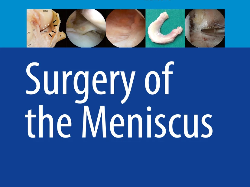
Abstract: The idiom ‘A picture is worth a thousand words’ refers to the notion that a complex idea can be conveyed with a single image more effectively then a description does. This chapter will focus on the astonishing meniscus anatomy. A number of highly detailed pictures are provided, highlighting the fact that an in-depth anatomical knowledge has a direct impact on meniscal reconstruction techniques (including any meniscal surgery: meniscectomy, suturing or meniscus replacement), thus determining their final clinical outcome.
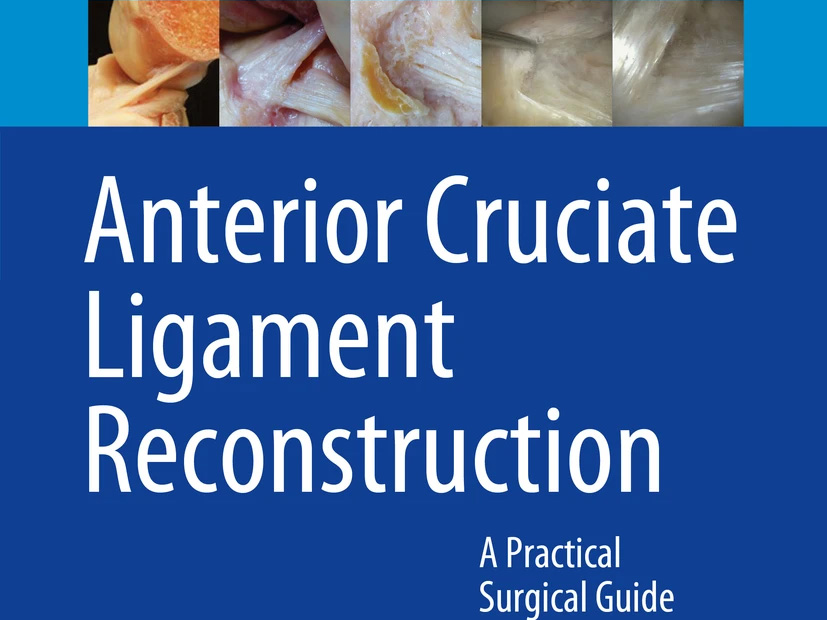
A Practical Surgical Guide
Abstract: An anatomical cadaveric study was performed in 111 knees to evaluate the macroscopic appearance of the tibial midsubstance of the ACL and its tibial insertion. The ACL was flat like a ribbon with a “C”-shaped tibial insertion along the medial tibial spine to the anterior aspect of the anterior horn of the lateral meniscus. Three variations of the tibial insertion were found. The bony insertion of the anterior horn of the lateral meniscus is in the center of the “C”-shaped tibial ACL insertion. Fibers of the anterior and posterior horn of the lateral meniscus blend with the tibial ACL insertion.

Prevention, Diagnosis, Treatment and Rehabilitation
Abstract: Groin pain and tenderness is one of the most frequent complaints among athletes; however, there is still a lot of confusion regarding the best treatment. In this chapter, we focus on some aspects of the most frequent and/or the most controversial pathologies in the groin area, among others: hip, osteitis pubis, adductor tendons, iliopsoas pathologies, different nerves entrapment or sport hernia. We also try to answer the question why there are so many different opinions regarding the best treatment.
Kilkadziesiąt publikacji w renomowanych międzynarodowych czasopismach naukowych takich jak; Annals of the Rheumatic Diseases, Knee Surgery, Sports Traumatology, Arthroscopy (KSSTA), The bone & joint journal i The Jurnal of Foot and Ankle Surgery.
Lorem ipsum dolor sit amet, consectetur adipiscing elit. Nullam eros felis, aliquet at placerat nec, aliquet eget quam. Nulla porttitor vel nunc sit amet finibus. Curabitur malesuada, turpis et rhoncus mollis, nisl nunc lobortis risus, sed placerat dui massa eget nisi. Fusce hendrerit eros vel convallis malesuada. Nullam mattis metus sit amet eros commodo, non euismod mi ultrices.

Lorem ipsum dolor sit amet, consectetur adipiscing elit. Nullam eros felis, aliquet at placerat nec, aliquet eget quam. Nulla porttitor vel nunc sit amet finibus. Curabitur malesuada, turpis et rhoncus mollis, nisl nunc lobortis risus, sed placerat dui massa eget nisi. Fusce hendrerit eros vel convallis malesuada. Nullam mattis metus sit amet eros commodo, non euismod mi ultrices.
Lorem ipsum dolor sit amet, consectetur adipiscing elit. Nullam eros felis, aliquet at placerat nec, aliquet eget quam. Nulla porttitor vel nunc sit amet finibus. Curabitur malesuada, turpis et rhoncus mollis, nisl nunc lobortis risus, sed placerat dui massa eget nisi. Fusce hendrerit eros vel convallis malesuada. Nullam mattis metus sit amet eros commodo, non euismod mi ultrices.
Kilkadziesiąt publikacji w renomowanych międzynarodowych czasopismach naukowych takich jak; Annals of the Rheumatic Diseases, Knee Surgery, Sports Traumatology, Arthroscopy (KSSTA), The bone & joint journal i The Jurnal of Foot and Ankle Surgery.
Autorzy: Robert Śmigielski, Urszula Zdanowicz, Michał Drwięga, Bogdan Ciszek, Beata Ciszkowska-Łysoń, Rainer Siebold
Abstract:
Purpose
Recently, the configuration of the anterior cruciate ligament (ACL) from its direct femoral insertion to midsubstance was found to be flat. This might have an important impact for anatomical ACL reconstruction. The purpose of this anatomical study was to evaluate the macroscopic appearance of the ACL from femoral to midsubstance.
Methods
The ACL was dissected in 111 human fresh frozen cadaver knees from its femoral insertion to midsubstance, and the shape was described. The anatomical findings were documented on digital photographs and on video. Thirty knees were sent for computed tomography (CT), magnetic resonance imaging (MRI) and histology of the femoral ACL insertion.
Results
Two millimetres from its direct femoral insertion, the ACL fibres formed a flat ribbon in all dissected knees without a clear separation between AM and PL bundles. The ribbon was in exact continuity of the posterior femoral cortex. The width of the ribbon was between 11.43 and 16.18 mm and the thickness of the ACL was only 2.54–3.38 mm. 3D CT, MRI and the histological examination confirmed above findings.
Conclusion
This is a detailed anatomical study describing the ribbon-like structure of the ACL from its femoral insertion to midsubstance. A key point was to carefully remove the surface fibrous membrane of the ACL. A total of 2–3 mm from its bony femoral insertion, the ACL formed a flat ribbon without a clear separation between AM and PL bundles. The ribbon was in exact continuity of the posterior femoral cortex. The findings of a flat ligament may change the future approach to femoral ACL footprint and midsubstance ACL reconstruction and to graft selection.
Zapisz się, jeżęli chcesz otrzymywać informacje o wartościowych szkoleniach, kursach kadawerowych oraz kluczowych publikacjach naukowych z zakresu ortopedii kończyny dolnej, medycyny sportowej i terapii regeneracyjnych.
Lekarz specjalistka w chirurgii ortopedycznej kolana, stopy i kostki. Członkini kadry naukowej McGowan Institute for Regenerative Medicine na Uniwersytecie w Pittsburghu i konsultant współpracujący ze związkami sportowymi i Polskim Komitetem Olimpijskim.
© 2025 Dr Urszula Zdanowicz. All rights reserved.
Polityka Prywatność